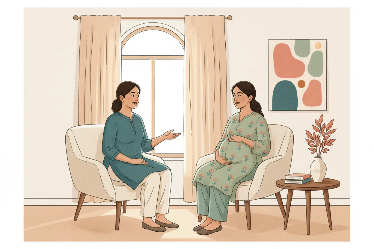

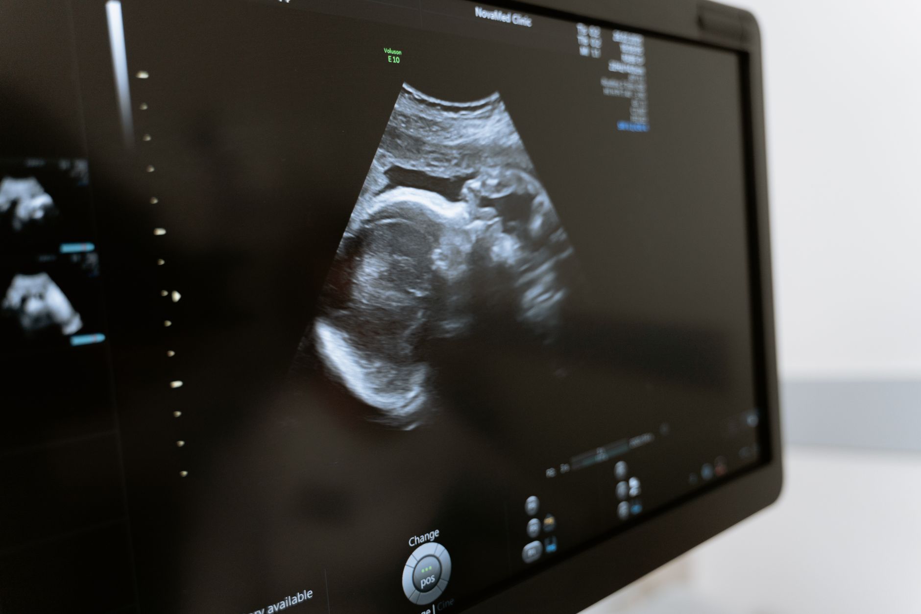
A growth scan in the last few weeks does not change your due date. If your pregnancy was already dated by an early scan, that date stands — later measurements should not change the gestational age or delivery date if a crown-rump length was already measured in the first trimester. A scan at 36 weeks answers a different question: how is this baby growing right now?
And the weight on that report is an estimate produced by a formula, not a measurement. In a large 2022 study of term scans, only about 71.6% of estimates came within 10% of the baby's actual birth weight.
Not medical advice — consult your obstetrician for guidance about your own pregnancy.
Why "The Scan Says 38 Weeks" Is Not a New Due Date
Dating is done once, as early as possible, and then left alone. First-trimester ultrasound, before 13 weeks and 6 days, is the most accurate method for establishing gestational age, at about 5 to 7 days, while a third-trimester scan is the least accurate dating method, at roughly 21 to 30 days. The same review is explicit that once a first-trimester scan has established or confirmed the estimated date of delivery, later findings should not be used to revise it.
| When the scan is done | What it is good for | Roughly how accurate for dating |
|---|---|---|
| Before 13 weeks 6 days | Setting the due date | About 5 to 7 days |
| At or after 28 weeks | Judging growth and wellbeing | About 21 to 30 days |
So "38 weeks 2 days by biometry" on a report at 36 weeks is not saying you are further along than you thought. It is saying this baby's head, abdomen and thigh bone currently measure like an average 38-week baby's. Babies at the same gestation are simply different sizes, and that spread is exactly why these late measurements should not change the gestational age or delivery date once a first-trimester crown-rump length exists.
In Indian private practice a third-trimester growth scan is close to routine, so parents now see a number that simply would not have existed a generation ago. It is a useful number. It is not a date.
What a Growth Scan Actually Measures
A growth scan takes measurements of the head, abdomen and femur — the thigh bone — which generate an estimated weight of your baby at that time, plus an estimate of the amniotic fluid around your baby and an assessment of the blood flow from the placenta to the baby through the umbilical artery. Those measurements go into a formula, and the formula produces a weight.
That formula has a published error bar, and it is wide. In a large 2022 study, the mean percent error between the estimated and actual birth weight was 2.39% plus or minus 9.13% — individual estimates scatter a long way either side of the truth even though the average sits close.
Put concretely: a 10% band around an estimate of 3.36 kg runs from roughly 3.02 kg to 3.70 kg, and nearly three in ten estimates land outside a band that wide. Accuracy is worse at the top end, where ultrasound's ability to predict birth weights over 4,500 g is imprecise, with sensitivity of only 33% to 44%.
"Big Baby" — What the Words Actually Mean
Two labels get used interchangeably, and they are not the same thing.
| Label | What it means | How it's judged |
|---|---|---|
| Large for gestational age (LGA) | Bigger than most babies at the same gestation | A centile on a growth chart |
| Macrosomia | Big in absolute terms, regardless of dates | A birth weight threshold |
Large for gestational age means an estimated fetal weight above the 90th customised centile, on a chart adjusted for the mother's height, weight in early pregnancy, parity and ethnic origin — which matters here, because a chart built on another population shifts where your baby appears to sit.
Macrosomia is the absolute-size label. Birth weights of 4,000 g or 4,500 g have historically been used to classify it, no universal definition has been accepted, and the most accurate way to diagnose it is to weigh the newborn after delivery — no single ultrasound, fundal height measurement or abdominal palpation can diagnose it alone.
What the Evidence Says About Acting on It
Start with what a late scan can and cannot predict. A meta-analysis of third-trimester ultrasound screening found an estimated fetal weight above 4,000 g detected only 53.2% of babies actually born above 4,000 g, and predicted shoulder dystocia with just 22% sensitivity — its authors urged caution before universal screening, because it would raise intervention rates without clear evidence of reducing newborn harm.
Guideline bodies read that same evidence cautiously. NICE's evidence review concluded the available evidence was not sufficient to recommend inducing labour, and that the risks and benefits of different modes of birth should be discussed with women with suspected fetal macrosomia — a conversation, not a protocol. The largest recent trial points the same way: the UK Big Baby trial randomised women whose babies were suspected to be large to induction at around 38 weeks or standard care, and found only a statistically non-significant reduction in shoulder dystocia, from 3.1% to 2.3%.
Caesarean is a separate question, and the thresholds discussed are high. ACOG, as summarised in the StatPearls macrosomia review, says an elective caesarean may be considered where the estimated fetal weight is above 5,000 g without gestational diabetes, or above 4,500 g with it — other bodies set that line differently, or set none. And most shoulder dystocias occur in mothers without diabetes carrying average-sized babies, while many macrosomic babies have none, which is why early induction for suspected macrosomia is not recommended on current evidence.
Blood Sugar Is Part of the Picture
When growth runs ahead of dates, blood sugar is one of the first things a clinician will want to look at. That is standard workup, not a diagnosis. India's national approach is to test every pregnant woman rather than only those with risk factors — the DIPSI test at booking and, if negative, repeated between 24 and 28 weeks — precisely because selective testing would mean missed cases.
What Goes Into the Delivery Decision
Estimated size is one input, and on its own the weakest. The growth scan reports more than a weight — amniotic fluid volume and umbilical artery blood flow are read alongside it. Your obstetric history, the baby's position and lie, findings on examination, and how labour actually starts and progresses all feed in, and none of those can be read off a 36-week report.
Which is why "wait a week and rescan" is an ordinary plan. A repeat scan shows the trend, and a trend beats a single reading taken with a method whose estimates land within 10% of the true birth weight only about 71.6% of the time.
The pressure in an Indian family often runs the other way. A number travels fast through a family WhatsApp group, and by evening someone has decided the baby is "too big" and a planned caesarean is the safe option. Worth keeping that conversation separate from the clinical one — the decision is yours and your obstetrician's, with the actual numbers in front of you.
Questions Worth Asking at Your Next Visit
- What date is my pregnancy dated from — my last period, or a first-trimester scan? Which is on my file?
- What centile is this estimated weight on, and which growth chart is that centile from?
- Apart from the estimated weight, what did the amniotic fluid and the Doppler show?
- What did my gestational diabetes test show, and does anything on this scan change whether I should be retested?
- If we rescan in a week, what change would alter your advice — and what would leave it unchanged?
- If size does become the main concern, what are my options, and what are the risks and benefits of each for me specifically?
Frequently Asked Questions
My scan says 38 weeks but my dates say 36. Which one is right?
Your dates, if they came from an early scan. Third-trimester measurements should not change the gestational age or delivery date when a first-trimester crown-rump length was already measured. The late scan describes size, not gestation.
Can the estimated weight really be off by a few hundred grams?
Yes, routinely. Only about 71.6% of estimates fall within 10% of the actual birth weight, with a mean percent error of 2.39% plus or minus 9.13%. At an estimate of 3.36 kg, a 10% band alone spans roughly 3.02 kg to 3.70 kg.
Does a big baby on the scan mean I need a caesarean?
Not by itself, and this is a decision for you and your obstetrician rather than something to settle from an article. The estimated weights at which an elective caesarean is even suggested for consideration are above 5,000 g without gestational diabetes, or above 4,500 g with it, and other bodies set the line differently or not at all.
Should I ask to be induced early, just in case?
Worth discussing, not assuming. NICE concluded the evidence was not sufficient to recommend inducing labour for suspected macrosomia, and that the risks and benefits of the different modes of birth should be discussed with the woman.
My mother-in-law is sure the baby is too big to deliver normally. What can I tell her?
That size alone predicts poorly. Most shoulder dystocias occur in mothers without diabetes carrying average-sized babies, while many macrosomic babies have none at all. Nobody can tell from a 36-week report how a particular labour will go.
Is repeating the scan in a week actually useful?
Often, yes — it shows a trend rather than a single point, and the scan also reports amniotic fluid volume and blood flow from the placenta to the baby, read alongside the weight. Ask what specifically would change the plan before the next scan is booked.